


An inguinal hernia is a defect in the groin region through which fat and/or intra-abdominal contents (such as the small intestine, colon, etc.) protrude. It has a wide range of clinical presentations, from a groin bulge detected during routine physical examination (with or without pain) to potentially life-threatening complications. These hernias can become complicated when they become incarcerated, meaning there is an acute mechanical bowel obstruction, or they may progress to strangulation, which increases the risk of perforation. Both of these situations are considered surgical emergencies. Elective (planned) surgical repair is the best treatment option for this condition.
The most common symptom associated with a hernia is a sensation of heaviness or discomfort in the groin area, which may or may not be associated with a visible bulge. Inguinal hernias may present as an intermittent swelling that depends on physical exertion. In some cases, this bulge shows slow growth, initially causing mild discomfort that later develops into intermittent pain. Groin discomfort is more pronounced when intra-abdominal pressure increases, such as when lifting heavy objects, straining, or standing for long periods of time.
Treatment is surgical in virtually all cases. There are selected cases of asymptomatic patients in whom surgery is not recommended due to associated comorbidities. This conservative approach with periodic monitoring is reserved for asymptomatic patients under strict medical supervision, particularly elderly patients with comorbidities who remain symptom-free. Inguinal hernia repair can be performed using different techniques. The two main approaches are open surgery and minimally invasive surgery.
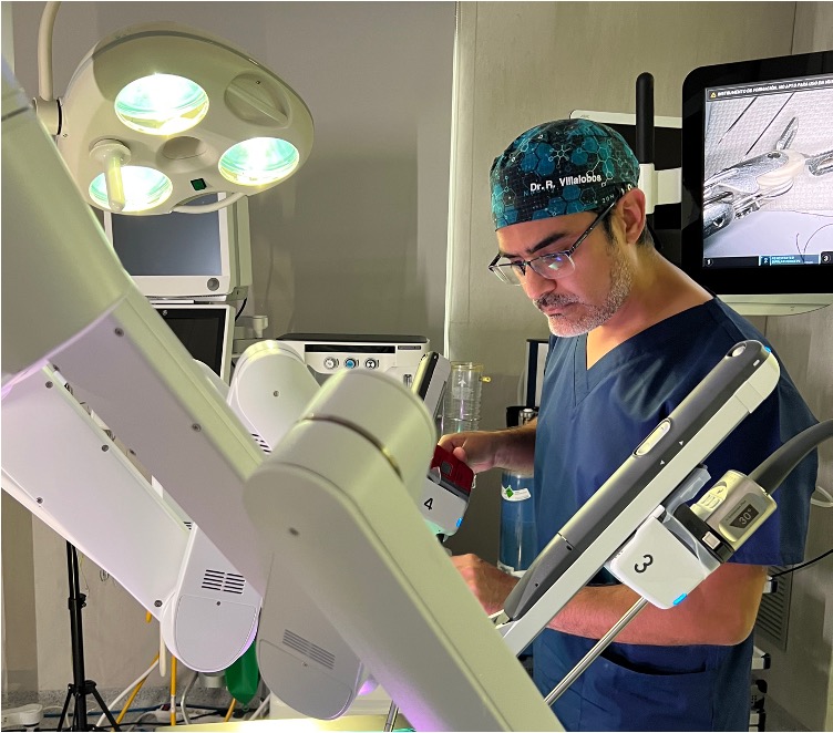
Open surgical repair involves a wide incision in the groin and dissection of the musculoaponeurotic structures of the abdominal wall, which then need to be sutured. Minimally invasive surgery (MIS) is performed via laparoscopy or robotics. In this case, MIS is carried out through three small ports, resulting in minimal damage to the abdominal wall. Robotic surgery provides an additional benefit of greater precision and improved visualization of the anatomy.
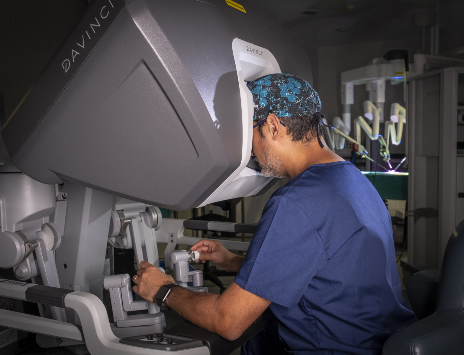
In all cases, the area is reinforced by placing a mesh, which may have different characteristics. Although all surgeons perform open inguinal hernia repairs, some also carry out laparoscopic repairs, and very few perform robotic surgery. Surgeons should choose the approach with which they feel most comfortable and have the greatest experience. For surgeons who are equally skilled in both techniques, the choice of surgical approach depends on the type of hernia and the patient’s characteristics.
Also called incisional hernias, as they are caused by previous surgical procedures. They are defects of the abdominal wall that can occur in any area depending on the location of the previous surgical scar. They are classified as midline, lateral, and posterior, and their treatment depends on the size of the defect and the volume of herniated contents, as well as the surgeon’s experience in using the most appropriate technique for each incisional hernia.
They are defects that can cause different types of symptoms, ranging from mild discomfort to frequent pain that limits daily activities. Even if they do not cause symptoms, treatment is recommended because almost 80% will eventually become symptomatic in some way, and some may develop complications requiring urgent surgical intervention. Therefore, elective surgical repair is recommended.
Treatment of incisional hernias can be performed in several ways, using either an open approach or a minimally invasive approach, i.e., laparoscopic or robotic surgery. Open surgery results in a more visible scar and has a longer recovery time compared to laparoscopic or robotic surgery.
Laparoscopic surgery has shown good results in experienced hands; however, it still has the disadvantage that suturing the defect is very difficult due to the surgeon’s limited ergonomics and discomfort during the procedure. Robotic surgery, on the other hand, has provided a major qualitative leap in the repair of this type of incisional hernia, with outcomes that in many cases surpass those of laparoscopy.
It is a separation greater than 2 cm between the rectus abdominis muscles at the level of the midline (linea alba), leaving a weakened and unprotected central area of the abdominal wall. The causes of rectus diastasis are varied. On one hand, there are congenital factors, although these are less common. On the other hand, there are cases related to aging and progressive weakening of the tissues, significant weight gain and increased abdominal volume, and most frequently, pregnancy. Approximately two-thirds of pregnant women develop rectus diastasis during the third trimester. This is understandable considering the significant abdominal distension that occurs during pregnancy. In addition, pregnancy is associated with increased levels of the hormone relaxin, which inhibits collagen formation and can lead to weakening of the connective tissue. If diastasis occurs only due to excessive stretching of the linea alba, it can usually return to its baseline state through supervised physical exercise guided by a professional. However, if the fibers of the linea alba are torn, it is more common for the midline separation to persist, and no amount of exercise will bring the rectus muscles back together. In these cases, the only way to restore the original anatomy is through surgery.
There is a variety of symptom presentations, but the most common are dissatisfaction with physical appearance and instability of the entire abdominal circumference, which can extend to the back. This is understood as an alteration of the abdominal core.
Treatment is highly variable, as there are techniques aimed at strengthening the abdominal musculature through specific physiotherapy. However, in many cases surgery is required to repair the separation. Surgical techniques vary depending on whether an open approach is used (i.e., with an incision) or a minimally invasive approach (laparoscopic or robotic). This surgery provides a definitive result and, in most cases, is reinforced with a mesh.
Chronic groin pain can have multiple causes. The most common are previous inguinal hernia surgery or athletic pubalgia (sports hernia), which is described in the next section. Persistent postoperative pain is defined as pain that does not improve, or even worsens, at least one month after inguinal hernia surgery. Fortunately, it occurs in less than 5% of patients, but in those who are affected it can significantly limit quality of life.
The patient presents variable symptoms, ranging from a constant discomfort to intense sharp pain during any physical activity, and in some cases even at rest. It is essential to have previously ruled out a hernia recurrence, meaning that the hernia has not reappeared.
Initial treatment consists of rest, anti-inflammatory medications, and anticonvulsants such as gabapentin or pregabalin. If there is no improvement, infiltrations with different types of medications (anesthetics and corticosteroids) may be performed. In cases where pain persists, surgery may be considered, in which the sensory nerves in the affected area are divided. Minimally invasive techniques (through small ports) have shown good results, although outcomes are not always positive.
Athletic pubalgia is known as chronic pain in the groin region, but it does not meet the criteria of a true hernia, as there is no actual anatomical defect. In this condition, no such defect is present. The most common causes include strain or injury to a muscle, tendon, or ligament, either in the lower limb (adductors) or in the abdominal wall (rectus abdominis, transversus abdominis, pyramidalis, external oblique, etc.). This condition often occurs in elite athletes such as football (soccer) players, hockey players, and American football players. This condition is sometimes referred to as a “sports hernia,” although the term is misleading, since it is not a true hernia as previously mentioned. Athletic pubalgia is part of a group of conditions affecting the groin region known as inguinal muscle imbalance syndrome. This problem occurs when there is a weak point in the abdominal muscle wall that allows internal organs to exert pressure through it. Surgery is required to correct this weak point.
The clinical presentation is very non-specific and variable, although the most common symptom is persistent groin pain, especially during physical activity. A thorough evaluation by multiple specialists (abdominal wall surgeon, orthopaedic surgeon, urologist, radiologist, and sports medicine physician) is required for an accurate diagnostic assessment.
Initial management consists of rest, anti-inflammatory medications, and rehabilitation therapy. Unfortunately, success rates are low, so surgical treatment is often required. There is no clear consensus on the most appropriate surgical technique; however, minimally invasive surgery (through small ports) has shown good results due to improved visualization of the entire groin region and reinforcement of the posterior wall with a mesh, given that a high percentage of patients present weakness in this area.
Our main values are humane treatment of the patient and professionalism to achieve excellence in all pathologies within our specialty.
Our main values are humane treatment of the patient and professionalism to achieve excellence in all pathologies within our specialty.

LEGAL INFORMATION
CONTACT
- info@cirugiaroboticaabdominal.com
-
Barcelona: TEKNON Medical Center
12 Marquesa de Vilallonga Street
Order 53
08017 Barcelona
Tel: +34 655754310
-
Zaragoza: Viamed Montecanal Hospital:
C. Franz Schubert, 2, 50012 Zaragoza.
Phone: +34 976218237
- Copyright 2023 Robotic Abdominal Surgery
